Warning: mkdir(): Permission denied in /home/virtual/lib/view_data.php on line 86 Warning: fopen(/home/virtual/tipjournal/journal/upload/ip_log/ip_log_2024-05.txt): failed to open stream: No such file or directory in /home/virtual/lib/view_data.php on line 88 Warning: fwrite() expects parameter 1 to be resource, boolean given in /home/virtual/lib/view_data.php on line 89 Endovascular Approach for Arterial Occlusion in Blunt Trauma: Two Cases
TIP Search

CLOSE


Trauma Image and Procedure > Volume 2(1); 2017 > Article
Kim, Kim, Cho, and Won: Endovascular Approach for Arterial Occlusion in Blunt Trauma: Two Cases

Abstract

Arterial occlusion after blunt trauma is rare, but can be devastating to the supplied organs or limbs. Management can vary from simple observation to surgery, such as thrombectomy or bypass, which may not be structurally possible and can inflict additional damage. Thus, an endovascular approach is another management option. We present two cases of arterial occlusion after blunt trauma, one successfully treated by endovascular procedure and one failure of the endovascular treatment.

CASE 1

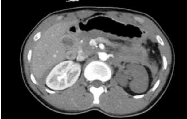
A 22-year-old male arrived at the emergency department complaining of abdominal pain. He was struck on the mid abdomen by a slow moving truck. He was alert and his vital signs were stable. Contrast-enhanced computed tomography (CT) of the abdomen demonstrated left renal artery occlusion along with total infarct of the left kidney (Fig. 1.). There was no gross hematuria. He was brought to the intervention room for renal angiography. Unfortunately, the artery was totally blocked, making passage of the guide wire impossible (Fig. 2.). Otherwise, there was no active bleeding, and he was observed. Follow-up CT performed 2 weeks later showed some collateral perfusion (Fig. 3.). Authors decided observation for the left kidney. The patient was discharged without complication.

CASE 2

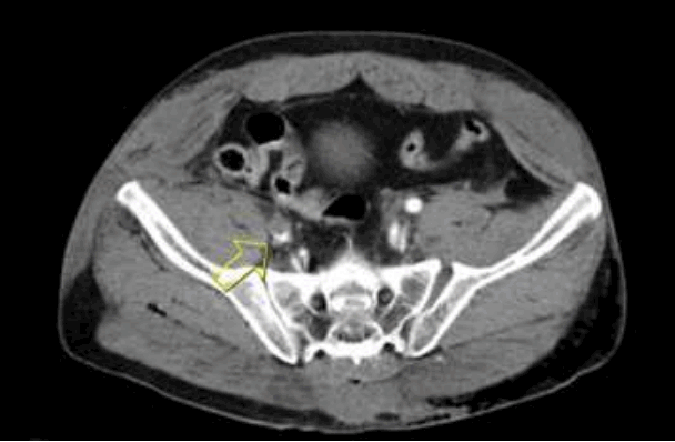
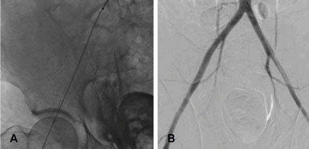
A 44-year-old male arrived at the emergency department complaining of pelvic and right leg pain after falling from a 2 m height. He demonstrated unstable vital signs, which were stabilized after crystalloid infusion. Physical examination revealed an unstable pelvis and weak right femoral pulse. Contrast-enhanced CT of the abdomen and pelvis demonstrated right inferior and superior rami fractures, left sacroiliac joint diastasis, and right external iliac arterial occlusion by thrombosis (Fig. 4.). Angiography revealed occlusion of the right external iliac artery, extending to the common femoral artery (Fig. 5.). The guide wire was able to pass and a stent graft was inserted. The post-stent angiography showed good arterial flow (Fig. 6.) and his femoral pulse returned to normal. His pelvis fracture was later managed by internal fixation, and 1 month later he was able to walk and was discharged without complication.

DISCUSSION

Arterial occlusions, especially those of peripheral arteries, after blunt trauma were reported 50 years ago [1]. Ischemia secondary to trauma can be devastating. There are several reports of arterial occlusions with various managements, such as patch angioplasty [2] and treatment via an endovascular approach [3]. Decisions for treatment may differ depending on the inflicted artery, type and force applied, and timing. Renal artery occlusion, for instance, may not always require surgical revascularization when there is a functioning contralateral kidney [4]. Trauma surgeons should be aware of various options, especially endovascular interventions when managing such patients.

Notes

Conflict of Interest Statement

No potential conflict of interest relevant to this article was reported.

Fig. 1.
Left renal artery occlusion
tip-2-1-22f1.gif
Fig. 2.
Angiography shows total obstruction of the left renal artery.
tip-2-1-22f2.gif
Fig. 3.
Follow-up CT (2 weeks later) shows some collateral perfusion.
tip-2-1-22f3.gif
Fig. 4.
CT shows left sacroiliac joint diastasis and right external iliac artery occlusion (arrow).
tip-2-1-22f4.gif
Fig. 5.
Angiography shows right external iliac artery occlusion by thrombosis, extending to common femoral artery.
tip-2-1-22f5.gif
Fig. 6.
(A) Guide wire was able to pass, and a 10-cm stent graft was inserted. (B) Post-stent angiography shows good arterial flow.
tip-2-1-22f6.gif

REFERENCES

1. Ophir M, Shulemson M, Laufer M, Sinkower A. Acute peripheral arterial occlusion due to blunt traumatic rupture of the intima. Israel journal of medical sciences. 1967;4(4):905-7.

2. Mogannam AC, Cubas RF, Gutierrez IM, Astudillo JA, Abou-Zamzam AM. Blunt Traumatic Occlusion of the Common Iliac Artery Repaired With Segmental Excision and Internal Iliac Artery Patch Angioplasty. Annals of vascular surgery. 2017;39:284 e1-. e4.
crossref
3. Mine T, Murata S, Yasui D, Tajima H, Kawamata H, Yokota H, et al. Endovascular recanalization techniques for popliteal arterial occlusive injury with limb-threatening ischemia secondary to trauma. Acta radiologica short reports. 2014;3(1):2047981613518772.
crossref pmid pmc
4. Santucci RA, Wessells H, Bartsch G, Descotes J, Heyns C, McAninch J, et al. Evaluation and management of renal injuries: consensus statement of the renal trauma subcommittee. BJU international. 2004;93(7):937-54.
crossref pmid


ABOUT
ARTICLE CATEGORY
Article Category

Browse all articles >

BROWSE ARTICLES
FOR AUTHORS AND REVIEWERS
Editorial Office
Korean Association for Research, Procedures and Education on Trauma
#1618, 18, Mapo-daero 4da-gil, Mapo-gu, Seoul 04177, Korea
Tel: +82-2-3280-9013    E-mail: karpet@karpet.or.kr

Copyright © 2024 by Korean Association for Research Procedures and Education on Trauma.

Developed in M2PI

Close layer
prev next